Search This Blog
Tuesday, June 26, 2012
Marfan Syndrome
Marfan syndrome is an inherited disorder of connective tissue, although about one-quarter of all cases occur without any family history of the syndrome. Marfan syndrome is caused by a defect in the fibrillin 1 gene on chromosome 15. It affects both men and women of all ethnic backgrounds. About 1 in 5,000 people have Marfan syndrome.
Symptoms
Marfan syndrome affects many areas of the body. Symptoms may include:
* tall and thin body type
* long arms, legs, fingers, and toes
* flexible joints
* curvature of the spine (scoliosis)
* chest sinks in (pectus excavatum) or sticks out (pectus carinatum)
* weak and fragile aorta, the main artery that carries blood away from the heart - it may become enlarged (aortic dilation) or develop a bulge (aneurysm), or may tear and burst (aortic dissection)
* severe nearsightedness, and sometimes dislocated lens of the eye
A person with Marfan syndrome has at least 3 symptoms in different parts of the body.
Diagnosis
If Marfan syndrome is suspected, a thorough physical examination should be done, as well as special testing. This would include:
* A complete family medical history and family tree
* Echocardiogram of the heart by a cardiologist
* Examination of the skeleton and measurement of body proportions
* Full eye examination by an eye doctor
Treatment
There is no cure for Marfan syndrome, but there are treatments that can help prevent or minimize its complications. The biggest threat to people with the syndrome is the sudden tearing of the aorta, a medical emergency which can be fatal. Therefore, close monitoring by a cardiologist and surgical repair of a weak aorta will save many lives. Blood pressure medications can also reduce stress on the aorta, as can avoiding strenuous exercise and contact sports.
In addition, regular physical examinations to monitor bones and joints and eye exams to watch for lens dislocation will help the person with Marfan syndrome. With good medical care most people can have a good quality of life and live a normal lifespan.
source: sangga or click here
Dandy-Walker Syndrome
Dandy-Walker Syndrome is a congenital condition affecting the cerebellum, an area in the lower back of the brain. Excess fluid in the areas surrounding the cerebellum impairs the brain's ability to control movement, and can also cause hydrocephalus and intracranial pressure. Other common features of the syndrome include an absence of the area between the hemispheres of the cerebellum, and a cyst at the base of the skull. It may be diagnosed at birth due to enlarged head size, or later in infancy or childhood when motor-skill problems appear. There is no cure, but treatment is available to lessen the impact of the symptoms.
Symptoms of Dandy-Walker Syndrome
Symptoms of Dandy-Walker syndrome vary according to age. Symptoms that appear in early infancy include: slow motor development and an abnormally enlarged skull. In older children symptoms include: irritability, vomiting, convulsions, ataxia and jerky eyes.
Other common symptoms of Dandy-Walker syndrome are:
* increased head circumference
* a head bulge at the back of the skull
* problems with the nerves that control the eyes, face and neck
* abnormal breathing patterns
Treatment
Treatments for Dandy-Walker syndrome are aimed at controlling and reducing the symptoms. In some cases, a special tube to reduce intracranial pressure may be placed inside the skull to control swelling.
source: sangga or click here
Saturday, June 23, 2012
Chediak-Higashi Syndrome

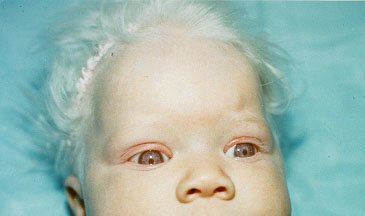
Chediak-Higashi syndrome (CHS) is characterized by partial oculocutaneous albinism (OCA), immunodeficiency, and a mild bleeding tendency. Approximately 85% of affected individuals develop the accelerated phase, a lymphoproliferative infiltration of the bone marrow and reticuloendothelial system. Adolescents and adults with atypical CHS and children with classic CHS who have successfully undergone allogenic hematopoietic stem cell transplantation (HSCT) develop neurologic findings during early adulthood.
Symptoms
Children with this condition may have:
* silver hair
* light colored eyes (albinism)
* jerky eye movements
* dicreased vision
* intellectual disability
* muscle weakness
* nerve problem in the limbs
* nosebleeds or easy bruising
* numbness
* tremor
* seizures
* sensitivity to light (photophobia)
Diagnosis
The diagnosis of Chediak-Higashi syndrome (CHS) should be considered in individuals with pigment dilution defects of the hair, skin, or eyes; congenital or transient neutropenia; immunodeficiency; and otherwise unexplained neurologic abnormalities or neurodegeneration. Each of these findings may be variably represented in affected individuals; therefore, heightened suspicion is needed to pursue an accurate diagnosis.
Treatment
There is no specific treatment for Chediak-Higashi syndrome. Bone marrow transplants appear to have been successful in several patients, especially when performed early in the disease.
Antibiotics are used to treat infections. Antiviral drugs such as acyclovir and chemotherapy drugs are often used in the accelerated phase of the disease. Surgery may be needed to drain abscesses in some cases.
source: sangga or click here
Friday, June 22, 2012
Maffucci Syndrome

Maffucci syndrome is a disorder which affects the skin and skeleton, causing benign cartilage tumors, bone deformities, and dark hemangiomas to develop. Despite these symptoms, individuals with Maffucci syndrome usually have a normal life span. Less than 200 cases of Maffucci syndrome have been published in the medical literature, so it is difficult to determine how often the syndrome occurs. The reports show it affects both males and females.
Symptoms
Maffucci syndrome has three main types of symptoms.
* Venous malformation (hemangioma) – These may be superficial or deep. If in the skin they often protrude as soft bluish bumps (nodules).
* Benign cartilage tumor (enchondroma) – These may appear anywhere in the body, but are most often found on the hands or feet, or long bones of the arms or legs. The enchondroma may cause the bone to weaken and break (pathologic fracture). About 30% of enchondromas may develop into cancerous tumors (chondrosarcomas).
* Bone deformities – This may include shortened length of the long bones, resulting in unequal arm or leg lengths. Bones may also break because they are weak, and when they heal, they may not align well (malunion).
Diagnosis
The symptoms of Maffucci syndrome seem to start in childhood, with parents often noticing hemangiomas of the skin in an affected child as early as age 4 or 5 years. The skin and bone growths develop slowly over time. Diagnosis of Maffucci syndrome is based on the symptoms present, since there is no specific laboratory test for the disorder.
Treatment
No medical care is needed for Maffucci syndrome. However, individuals affected by the syndrome need to have regular physical examinations to evaluate changes in the skin and bone lesions. In particular, health care providers should watch for changes that signal the development of a malignant tumor (chondrosarcoma). An orthopedic surgeon will evaluate bone changes and pathologic fractures, and a dermatologist will evaluate skin hemangiomas.
source: click here
Wednesday, June 20, 2012
Sjogren's Syndrome

Sjogren's syndrome is a condition which is often misdiagnosed or undiagnosed. Awareness and recognition of Sjogren's syndrome is important.
Experts believe 1 to 4 million people have the disease. Of this group, 90% are women. Sjogren's syndrome can occur at any age, but it usually is diagnosed after age 40 and can affect people of all races and ethnic backgrounds. Sjogren's syndrome is rare in children, but it can occur.
Many women have the disease but do not recognize the symptoms which are similar to those of other conditions including menopause.
Symptoms
It has been suggested that many more people suffer with Sjogren's syndrome than have been diagnosed. Sjogren's syndrome is considered to be:
* an autoimmune disease
* a rheumatic disease
* a connective tissue disorder
Abnormal production of autoantibodies in the blood which turn against various tissues in the body cause the disease. Inflammation in the glands of the body results from this abnormality.
Sjogren's syndrome is characterized by:
* dry mouth
* dry eyes
* hoarseness
* vaginal dryness
* skin dryness
* chronic fatigue
Diagnosis
The diagnosis of Sjogren's syndrome is based largely on the detection of dry eyes and mouth. Helpful diagnostic tools that serve in making the diagnosis include:
* Schirmer's test for dry eyes
* radiologic salivary scans
* salivary flow testing
* biopsy of salivary glands
* blood tests
Treatment
Treatment of Sjogren's syndrome is different for each person, depending on what parts of the body are affected. Although there is no cure for Sjogren's syndrome, mouthwashes, saliva substitutes, sprays, gels, and gum can relieve oral symptoms. Medications and drug treatment options for dry mouth associated with Sjogren's syndrome may include saliva and mucus stimulating drugs such as:
* Salagen (Pilocarpine hydrochloride)
* Evoxac (Cevimeline HCI)
Artificial tears and eye ointments can help relieve chronic dry eyes. Medications and drug treatment options for dry eye associated with Sjogren's syndrome may include:
* Restasis (Cyclosporine Opthalmalic Emulsion)
* Hydroxypropyl Cellulose (Eye Drops & Pellets)
Extraglandular problems, such as joint pain or muscle pain involvement, are often treated with NSAIDs (non-steroidal anti-inflammatory drugs). Lung, kidney, blood vessel, or nervous system problems may be treated with:
* corticosteroids
* DMARDs (disease-modifying anti-rheumatic drugs) or immunosuppressives
source: sangga or click here
Patau Syndrome (Trisomy 13) Extra copy of chromosome 13

Patau syndrome, Trisomy 13, is the least common of the autosomal trisomies, after Down syndrome (Trisomy 21) and Edwards syndrome (Trisomy 18). The extra copy of chromosome 13 in Patau syndrome causes severe neurological and heart defects which make it difficult for infants to survive. The exact incidence of Patau syndrome is not known, although it appears to affect females more than males, most likely because male fetuses do not survive until birth. Patau syndrome, like Down syndrome, is associated with increased age of the mother. It may affect individuals of all ethnic backgrounds.
Newborns with Patau syndrome share common physical characteristics:
* Extra fingers or toes (polydactyly)
* Deformed feet, known as rocker-bottom feet
* Neurological problems such as small head (microcephaly), failure of the brain to divide into halves during gestation (holoprosencephaly), severe mental deficiency
* Facial defects such as small eyes (microphthalmia), absent or malformed nose, cleft lip and/or cleft palate
* Heart defects (80% of individuals)
* Kidney defects
Diagnosis
The symptoms of Patau syndrome are evident at birth. Patau syndrome may be mistaken for Edwards syndrome, so genetic testing should be done to confirm the diagnosis. Imaging studies such as computed tomography (CT) or magnetic resonance imaging (MRI) should be done to look for brain, heart, and kidney defects. An ultrasound of the heart (echocardiogram) should be done given the high frequency of heart defects associated with Patau syndrome.
Treatment
Treatment of Patau syndrome focuses on the particular physical problems with which each child is born. Many infants have difficulty surviving the first few days or weeks due to severe neurological problems or complex heart defects. Surgery may be necessary to repair heart defects or cleft lip and cleft palate. Physical, occupational, and speech therapy will help individuals with Patau syndrome reach their full developmental potential
source: sangga or click here
Sunday, January 1, 2012
Juvenile Retinoschisis
Causes progressive loss of vision.
X-linked juvenile retinoschisis is a genetic disorder of the X chromosome. A gene identified as being responsible for juvenile retinoschisis, RS1, encodes an amino acid protein, retinoschisin, which is important in photoreceptor cells of the eye. Physical changes occur in the retina, the part of the eye responsible for vision. The retina splits into two layers, which impairs vision.
Since juvenile retinoschisis is an X-linked recessive disorder, it occurs primarily in boys (since they have only one X chromosome), but it can occur in females with two defective genes (one on each of their two X chromosomes). Juvenile retinoschisis occurs in individuals of all ethnic backgrounds.
Symptoms
Children with juvenile retinoschisis have gradually decreasing vision due to splitting of the retina. Physical changes in the eye may include:
* development of cysts and ruptured blood vessels between the layers of the retina
* holes in the retinal layers, which may lead to detachment of the retina from its underlying tissue (5-22% of individuals)
* leakage of blood into the jelly-like material inside the eye (vitreous hemorrhage)
* changes in the macula, the area of clearest vision in the retina.
Individuals with juvenile retinoschisis may also have difficulty focusing on an object (strabismus) and roving, involuntary eye movements (nystagmus).
Diagnosis
A thorough ophthalmological evaluation can help distinguish juvenile retinoschisis from similar retinal degenerative diseases such as retinitis pigmentosa. An imaging technique called optical coherence tomography (OCT) provides high-resolution cross-sectional images of the macula to look for abnormalities. OCT can show the changes present in juvenile retinoschisis. An electroretinogram will show dysfunction throughout the retina. Genetic testing can reveal the presence of the defective RS1 gene.
Treatment
No treatment is yet available to stop the progression of juvenile retinoschisis. Surgery can repair vitreous hemorrhage and retinal detachments. Low-vision aids, mobility training, and adaptive training skills can help individuals with vision loss. Genetic counseling can help identify family members who are carriers of the RS1 gene.
source: click here
Subscribe to:
Posts (Atom)